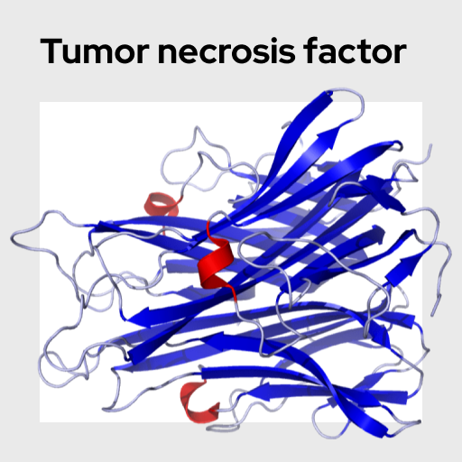
Tumor necrosis factor (TNF-α)
Tumor necrosis factor α (TNF-α) is a pleiotropic, proinflammatory mediator that is involved in a wide range of inflammatory, infectious, autoimmune and malignant conditions. This cytokine has been implicated in a variety of diseases, including autoimmune diseases, insulin resistance, and cancer. TNF-α has been shown to impact nervous system activity and may play a mechanistic role in the relationship between inflammation and behavior. In addition to increasing in response to infection, levels of this cytokine also increase in response to psychosocial stress. This analyte is eligible for multiplexing.
Name: Tumor necrosis factor alpha (TNF-α)
Category: Health & Inflammation
Type of test: Blood + Saliva
Tumor necrosis factor alpha (TNF-α) is a proinflammatory cytokine implicated in a large number of biological processes including, but not limited to, regulating apoptotic cell death, fever production, generating inflammation, anti-tumor activity, and the etiology of sepsis. All of the members of the TNF superfamily can be characterized as type II transmembrane proteins, whose activation is dependent on cleavage and subsequent release of the highly conserved TNF domain. As one might expect from something as multifaceted as TNF, it has been shown to be involved in a huge number of human pathologies. Dysregulation of TNF-α is linked to Alzheimer’s disease, cancer, depression, inflammatory bowel disease, rheumatoid arthritis, asthma, psoriasis, auto-immune disorders, and many others. Neutralization of this cytokine using antibodies is used as a treatment for several inflammatory disorders.
TNF-α is among the first signaling proteins to be released in the innate immune response to pathogen detection, and once present in the plasma it has numerous mechanisms of action. Within the liver, TNF binds and stimulates the production and subsequent release of acute phase proteins such as C-reactive protein (CRP). At sites of infection, it leads to microvascular hyperpermeability as well as vasodilation, which increases the ability of neutrophils and monocytes, as well as other immune cells, to infiltrate the target tissue. Once localized, macrophages are the primary cells to secrete TNF-α, alongside NK cells and T lymphocytes. The mechanism for its secretion involves ER synthesis and SNARE protein mediated fusion of TNF-α -vesicles with VAMP3 recycling endosomes. Once within the endosomes, a complex of proteins facilitates the translocation of TNF proteins towards the cell membrane. Rho1 and Cdc42 are two proteins involved in the remodeling of actin to allow for this movement towards the membrane, and once within the outer membrane the transmembrane proteins await cleavage by the enzyme TACE to enter their active soluble form.
TNF-α also binds to receptors in the hypothalamus and causes the synthesis and release of corticotropin-releasing hormone (CRH), triggering the production of cortisol and body's stress response. Furthermore, psychological stress leads to increases in TNF-α concentrations, making it a potentially valuable marker for the study of stress. TNF-α can be measured in both saliva and serum / plasma, but the extent to which levels in these sample types correlate is variable.
Parameswaran, N. & Patial, S. (2010). Tumor necrosis factor-α signaling in macrophages. Critical Review of Eukaryotic Gene Expression, 20, 87-103. https://doi.org/10.1615/critreveukargeneexpr.v20.i2.10
Slavish, D. C., Graham-Engeland, J. E., Smyth, J. M., & Engeland, C. G. (2015). Salivary markers of inflammation in response to acute stress. Brain, Behavior, and Immunity, 44, 253-269. https://pubmed.ncbi.nlm.nih.gov/25205395/
Steptoe, A., Owen, N., Kunz-Ebrecht, S., & Mohamed-Ali, V. (2002). Inflammatory cytokines, socioeconomic status, and acute stress responsivity. Brain, Behavior, and Immunity, 16, 774-784. https://psycnet.apa.org/record/2003-01040-006


