Insulin
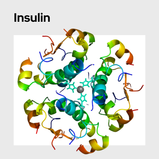
Insulin is an anabolic hormone produced by the pancreas. It promotes glucose uptake and metabolism in liver, fat, and muscle cells. Thus, increases in insulin levels lead to decreases in circulating blood glucose. Absent and dysregulated insulin signaling are at the core of Type I and Type II diabetes, respectively.
Name: Insulin
Category: Hunger and Satiety
Type of Test: Blood + Saliva
Insulin, an essential peptide hormone, is considered one of the oldest, most highly conserved proteins found in humans. As a peptide hormone, insulin is composed of 51 amino acids in a precisely folded chain. Insulin is considered to be the body’s primary anabolic hormone as it promotes the synthesis of proteins in variety of different tissues. Broadly, insulin is responsible for regulating the metabolism of the three main macromolecules: carbohydrates, fats, and proteins, by promoting the uptake of glucose into muscle, fat, and liver cells. Additionally, insulin also influences immune responses, growth, mitogenesis, fat storage, reproduction, cognition, lifespan, and cancer. However, insulin is most commonly known for its role in blood glucose regulation and its clinical significance to diabetes mellitus.
Insulin is generated in the beta cells (ß cells) of the pancreatic islets. Pancreatic beta cells are sensitive to blood glucose levels and the release of insulin peaks after each meal with small oscillations throughout the day. After release, insulin travels through the bloodstream. It acts on insulin receptors (IR) kinases, located in cell membranes in target organs, such as the liver, skeletal muscles, and adipose tissues. In the liver, insulin promotes the uptake of glucose and glucose storage in the form of glycogen, while it decreases glucose release. In the skeletal muscles and adipose tissues, insulin stimulates glucose transport to other tissues. Besides glucose metabolism, insulin can act on most cell types, including brain cells, T cells, and B cells.
Changes in levels of insulin have been linked to eating behavior and the subjective feelings of hunger and satiety. Specifically, low plasma insulin concentrations are associated with hunger, and insulin spikes after eating coincide with satiety. Additionally, insulin activity in the brain has been linked to changes in functional connectivity of regions associated with cognitive and homeostatic control of energy balance, suggesting that insulin plays a critical role in regulating eating behavior and weight changes. Moreover, insulin resistance is a characteristic feature of obesity and diabetes mellitus type II.
Insulin levels can be tested via serum / plasma or saliva sampling. While serum / plasma is often preferred for accurate representation of circulating levels of this hormone, salivary measures can be used as a minimally invasive method. Salivary insulin levels typically correlate positively with serum / plasma levels. Depending on the type of study, insulin should be tested after a three-hour fasting period (when levels are low) or directly after food intake (when levels spike).
Fekete, Z., Korec, R., Feketeova, E., Murty, V. L., Piotrowski, J., Slomiany, A., & Slomiany, B. L. (1993). Salivary and plasma insulin levels in man. Biochemistry and molecular biology international, 30, 623–629. https://pubmed.ncbi.nlm.nih.gov/8401319/
Huang, Z., Huang, L., Waters, M. J., & Chen, C. (2020). Insulin and Growth Hormone Balance: Implications for Obesity. Trends in Endocrinology & Metabolism, 31, 642-654. https://doi.org/10.1016/j.tem.2020.04.005
Kullmann, S., Heni, M., Veit, R. et al. (2017). Intranasal insulin enhances brain functional connectivity mediating the relationship between adiposity and subjective feeling of hunger. Scientific Reports, 7, 1627. https://doi.org/10.1038/s41598-017-01907-w
Taouis, M., & Torres-Aleman, I. (2019). Editorial: Insulin and The Brain. Frontiers in Endocrinology, 10, e00299. https://doi.org/10.3389/fendo.2019.00299


